Dosya:Apikal4D.gif
Apikal4D.gif (636 × 432 piksel, dosya boyutu: 705 KB, MIME türü: image/gif, döngüye girdi, 15 kare, 0,6 sn)
Bu dosya Wikimedia Commons deposunda bulunmaktadır ve diğer projeler tarafından kullanılıyor olabilir. Aşağıda dosya açıklama sayfasındaki açıklama gösteriliyor.
Özet
| AçıklamaApikal4D.gif |
العربية: صُورةٌ مُتحرِّكة لِتخطيط صدى القلب؛ دائرةٌ كaهربائيَّة ثُلاثيَّة الأبعاد للقلب كما يبدو للناظر من الأعلى، حيثُ تمَّت إزالة الجُزء القمّي من
البطينين بينما يظهر الصمَّام التاجي واضحًا للعيان. لا يظهر الصمَّامان الأبهر وثُلاثيّ الشرف بشكلٍ واضح نتيجة ضياع البيانات الخاصَّة بهذا التصوير، بينما تظهر الفتحات. يظهرُ على اليسار منظران ثُنائيَّا الأبعاد مأخوذان بناءً على بيانات الرسم ثُلاثي الأبعاد. يُفسِّرُ هذا الرسم الصُورة المُتحرِّكة.
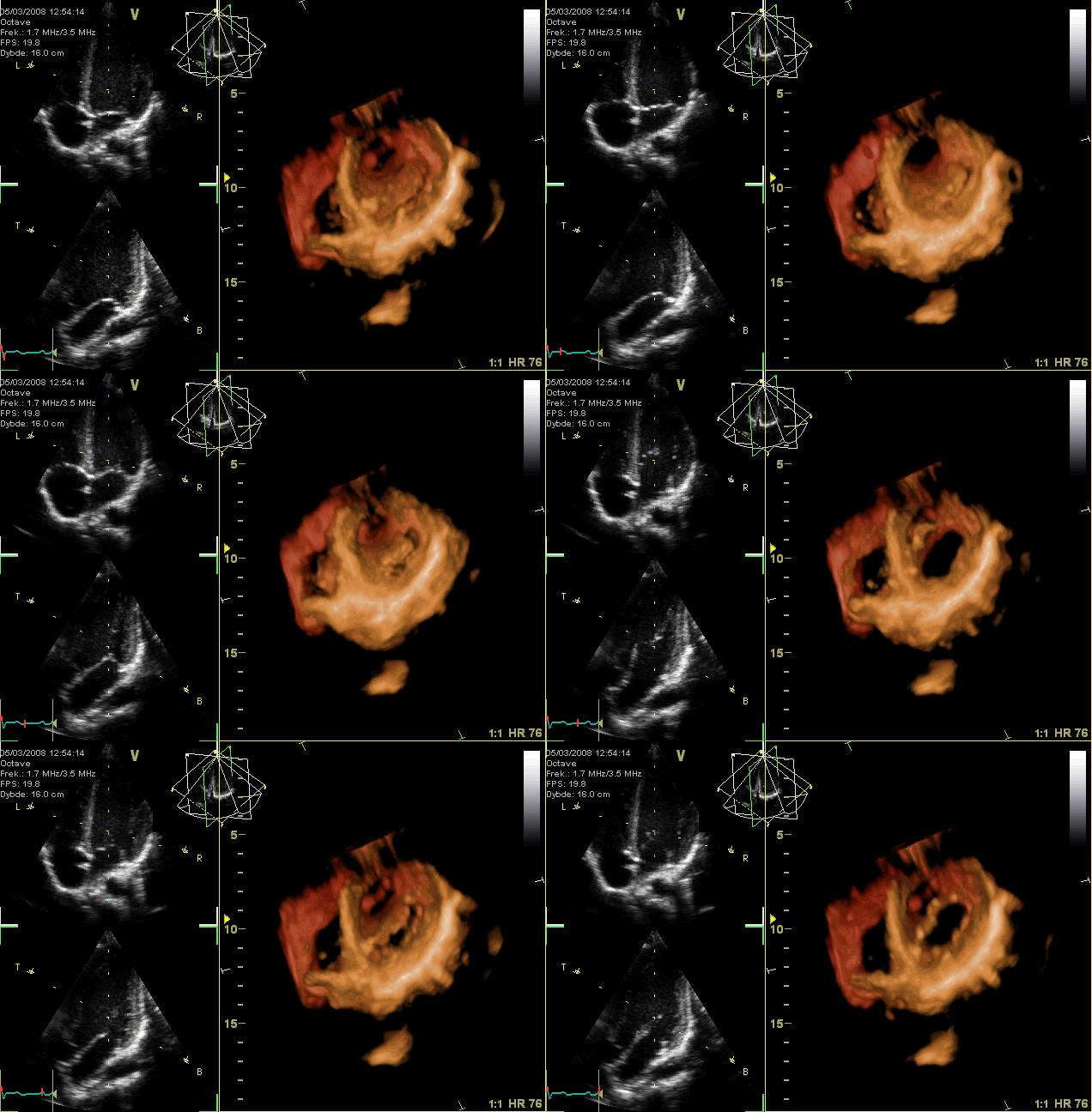
English: GIF-animation showing a moving echocardiogram; a 3D-loop of a heart viewed from the apex, with the apical part of the ventricles removed and the mitral valve clearly visible. Due to missing data the leaflet of the tricuspid and aortic valve is not clearly visible, but the openings are. To the left are two standard two-dimensional views taken from the 3D dataset.
Why might echocardiogram imaging be helpful? An echocardiogram is a non-invasive procedure used to evaluate the functionality and structure of the heart. This kind of imaging might be helpful as it allows the examination of the heart's anatomy and the blood vessels around it (NHS, 2022). The procedure also analyses how blood flows through the veins and evaluates the heart's pumping chambers. In addition, it aids in diagnosing and monitoring some cardiac diseases. What kinds of issues or anomalies might an echocardiogram imaging be able to detect? An echocardiogram might detect atherosclerosis, where fatty molecules and other substances in the bloodstream gradually block the arteries. This heart condition may result in issues with the heart's pumping or wall movements (Johns Hopkins Medicine, 2022). The procedure can also detect cardiomyopathy, a cardiac enlargement brought on by thick and frail heart muscle. Additionally, congenital heart defects that appear in one or more cardiac components while a fetus is still developing might be detected using this procedure (Hopkins Medicine, 2022). Moreover, failure of the heart might be noticed. This condition happens when blood cannot be pumped effectively because the heart muscle has grown weak or tight due to cardiac relaxation. Some symptoms of this condition include swelling in the feet, ankles, and other areas of the body and fluid accumulation in blood vessels and lungs. Furthermore, aneurysm and heart valve malfunction might be detected. An aneurysm is an enlargement and weakening of the aorta or a portion of the heart muscle. If this condition occurs, there is a chance that the aneurysm will burst. On the other hand, heart valve malfunction involves the failure of one or more heart valves, which could result in irregular blood flow within the heart. The constriction of the valves prevents adequate blood flow. As a result, blood might ooze backward through the defective valves, which is dangerous (Johns Hopkins Medicine, 2022). Therefore, using an echocardiogram, heart valves can be examined for infection. At the same time, doctors can determine the best treatment for these diseases. https://www.intechopen.com/chapters/44904 What might an echocardiogram not be able to detect? An echocardiogram cannot reveal if one has blocked or clogged arteries. According to Discoverecho (2019), echocardiography is not a medical test particularly good at finding blocked arteries. This setback is mainly due to coronary arteries being typically too tiny to be seen using echocardiography. References Discoverecho. (2019, Nov 13). Does an echocardiogram show blockages? (Blocked arteries). https://discoverecho.com/does-echocardiogram-show-blockages/ Jamil, G., Abbas, A., Shehab, A., & Qureshi, A. (2013, Jun 12). Echocardiography findings in common primary and secondary cardiomyopathies. https://www.intechopen.com/chapters/44904 Johns Hopkins Medicine. (2022). Echocardiogram. https://www.hopkinsmedicine.org/health/treatment-tests-and-therapies/echocardiogram NHS. (2022, Mar 28). Echocardiogram. https://www.nhs.uk/conditions/echocardiogram/ A Sketch explains the animation.Français : Animation GIF en boucle montrant une échocardiographie en trois dimensions animée. Le cœur est ici vu depuis son apex, avec la partie apicale des ventricules enlevée. La valve mitrale est clairement visible. A cause de la résolution, les feuillets des valves tricuspide et aortique ne sont pas visibles. A gauche se trouvent deux vues bidimensionnelles standard extraites des données tridimensionnelles. L'animation est expliquée par un schéma visible ici. |
| Tarih | |
| Kaynak | Yükleyenin kendi çalışması |
| Yazar | Kjetil Lenes |
| Diğer sürümler |
|
Değerlendirme
|
| This image was selected as a picture of the week on the Persian Wikipedia for the 46. week, 2010. |

|
This image was selected as picture of the day on Bengali Wikipedia.
|

|
Bu görüntü 4 Ocak 2011 tarihinde günün resmi olarak seçilmiştir. Görüntünün başlığı o tarihte aşağıdaki gibiydi: English: GIF-animation showing a moving echocardiogram; a 3D-loop of a heart viewed from the apex, with the apical part of the ventricles removed and the mitral valve clearly visible. Due to missing data the leaflet of the tricuspid and aortic valve is not clearly visible, but the openings are. To the left are two standard two-dimensional views taken from the 3D dataset. Diğer diller:
Dansk: GIF-animation af et ekkokardiogram; et 3-D-loop af et hjerte set fra apex, hvor apexdelen af ventriklerne er fjernet, og mitralklappen er tydeligt synlig. Manglende data betyder, at trikuspidal- og aortaklappen ikke ses tydeligt, men åbningerne gør. Til venstre ses to standard to-dimensionale billeder, udtaget fra 3-D dataene. English: GIF-animation showing a moving echocardiogram; a 3D-loop of a heart viewed from the apex, with the apical part of the ventricles removed and the mitral valve clearly visible. Due to missing data the leaflet of the tricuspid and aortic valve is not clearly visible, but the openings are. To the left are two standard two-dimensional views taken from the 3D dataset. Español: Animación de un ecocardiograma: ciclo 3D de un corazón visto desde arriba, con la parte apical de los ventrículos retirada y la válvula mitral claramente visible. Debido a la falta de datos la valva de las válvulas tricúspide y aórtica no es claramente visible, pero las aperturas sí. A la izquierda hay dos vistas estándares bidimensionales tomadas del conjunto de datos tridimensional. Interlingua: Animation GIF de un echocardiogramma; un representation 3D de un corde vidite ab le apice, con le parte apical del ventriculos removite e le valvula mitral clarmente visibile. Per manco de datos, le folietto del valvula tricuspide e aortic non es clarmente visibile, ma le aperturas lo es. Al sinistra il ha duo vistas standard bidimensional prendite del collection de datos 3D. Italiano: it:Ecocardiogramma del cuore in vista apicale 3D: la parte superiore del ventricolo è stata rimossa per mettere in evidenza la it:valvola mitrale. A sinistra sono riportate per confronto due convenzionali immagini bidimensionali. Magyar: GIF-animáció, amelyen egy szívultrahang mozgóképe (echokardiogramja) látható; térhatású ismétlődő felvétel a szívcsúcs felőli nézetben, elhagyva a kamrák csúcshoz tartozó részét, de láthatóvá téve a mitrális szívbillentyűt. Az adatok hiánya miatt a trikuszpidális és az aortabillentyű nem látszik jól, de a nyílásuk igen. Balra két kétdimenziós ábra szerepel a 3D-s adathalmazból kiemelve (a fenti képen a trikuszpidális és a mitrális billentyűk, az alsón az aorta- és a mitrális billentyűk ismerhetők fel). Nederlands: GIF-animatie van een bewegend echocardiogram; een 3D-loop van een hart gezien vanaf de apex (punt van het hart) met de mitralisklep duidelijk zichtbaar en zonder het apicale deel van de ventrikels (hartkamers). Door ontbrekende gegevens is het slipje van de tricuspidalis- en aortaklep niet duidelijk zichtbaar, maar de openingen ervan wel. Links twee standaard tweedimensionale aanzichten die aan de 3D-dataset zijn ontleend. Українська: GIF-анімація, що демонструє рухому ехокардіограму — тривимірне циклічне (3D-loop) зображення серця зверху. Верхівкова частина шлуночків видалена і мітральний клапан чітко видно. Через відсутність даних стулки трикуспідального (тристулкового) клапану і аортальний клапан не видно чітко, але отвори відкриті. Зліва два стандартні двовимірні зображення отримані з 3D даних. 日本語: GIFアニメーションによる心臓超音波検査の三次元動画。心臓を心尖側から見ている。両心室の心尖部が取り除かれ、僧帽弁がはっきりと見える。データ欠損のため三尖弁と大動脈弁の弁尖は鮮明には見えないが、その開口部は鮮明である。左側の2つの図は三次元データから作成された標準的な二次元画像である。 中文: 右侧为三维超声心动图,左侧为标准二维超声心动图。 |
Lisanslama

|
Bu belgenin GNU Özgür Belgeleme Lisansı, Sürüm 1.2 veya Özgür Yazılım Vakfı tarafından yayımlanan sonraki herhangi bir sürüm şartları altında bu belgenin kopyalanması, dağıtılması ve/veya değiştirilmesi için izin verilmiştir;
Değişmeyen Bölümler, Ön Kapak Metinleri ve Arka Kapak Metinleri yoktur. Lisansın bir kopyası GNU Özgür Belgeleme Lisansı sayfasında yer almaktadır.http://www.gnu.org/copyleft/fdl.htmlGFDLGNU Free Documentation Licensetruetrue |
- Şu seçeneklerde özgürsünüz:
- paylaşım – eser paylaşımı, dağıtımı ve iletimi
- içeriği değiştirip uyarlama – eser adaptasyonu
- Aşağıdaki koşullar geçerli olacaktır:
- atıf – Esere yazar veya lisans sahibi tarafından belirtilen (ancak sizi ya da eseri kullanımınızı desteklediklerini ileri sürmeyecek bir) şekilde atıfta bulunmalısınız.
- benzer paylaşım – Maddeyi yeniden karıştırır, dönüştürür veya inşa ederseniz, katkılarınızı orijinal olarak aynı veya uyumlu lisans altında dağıtmanız gerekir.
Altyazılar
Bu dosyada gösterilen öğeler
betimlenen
Vikiveri ögesi olmayan bir değer
5 Mart 2008
image/gif
a237b56e924fbd0eae3c097b3c87bce1e71d0ae5
721.588 bayt
0,6 saniye
432 piksel
636 piksel
Dosya geçmişi
Dosyanın herhangi bir zamandaki hâli için ilgili tarih/saat kısmına tıklayın.
| Tarih/Saat | Küçük resim | Boyutlar | Kullanıcı | Yorum | |
|---|---|---|---|---|---|
| güncel | 20.42, 13 Mart 2008 |  | 636 × 432 (705 KB) | wikimediacommons>Ekko | {{Information |Description=GIF-animation showing a moving echocardiogram; a 3D-loop of a heart wieved from the apex, with the apical part of the ventricles removed and the mitral valve clearly visible. Due to missing data the leaflet of the tricuspid and |
Dosya kullanımı
Aşağıdaki sayfa bu dosyayı kullanmaktadır:


