Dosya:Nematocyst discharge.png
Daha yüksek çözünürlük yok.
Nematocyst_discharge.png (480 × 371 piksel, dosya boyutu: 190 KB, MIME türü: image/png)

|
Bu galeride bulunan tüm resimler vektörel grafikler kullanılarak SVG dosyası şeklinde oluşturulmalıdır. Bunun çeşitli avantajları vardır; daha fazla bilgi için Commons:Temizleme medyası sayfasına bakın. Eğer bu resmin SVG formatına sahipseniz, lütfen yükleyin. Lütfen SVG dosyanızı yükledikten sonra, bu şablonu bu resimdeki {{vector version available|yeni resim ismi.svg}} şablonu ile değiştirin.
|
Özet
| Açıklama |
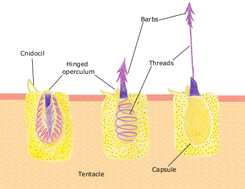
English: The diagram above shows the anatomy of a nematocyst cell and its “firing” sequence, from left to right. On the far left is a nematocyst inside its cellular capsule. The cell’s thread is coiled under pressure and wrapped around a stinging barb. When potential prey makes contact with the tentacles of a polyp, the nematocyst cell is stimulated. This causes a flap of tissue covering the nematocyst—the operculum—to fly open. The middle image shows the open operculum, the rapidly uncoiling thread and the emerging barb. On the far right is the fully extended cell. The barbs at the end of the nematocyst are designed to stick into the polyp’s victim and inject a poisonous liquid. When subdued, the polyp’s tentacles move the prey toward its mouth and the nematocysts recoil back into their capsules. |
| Tarih | 11 Nisan 2007 (original upload date) |
| Kaynak | en.wikipedia üzerinden Commons'a transfer edildi. |
| Yazar | The original uploader was Spaully at İngilizce Vikipedi. |
Lisanslama
This file is licensed under Creative Commons ShareAlike 1.0 License.
Creative Commons has retired this legal tool and does not recommend that it be applied to works.
|
Nematocyst discharge.png adlı görüntü kamu malıdır çünkü içeriği, ABD Ulusal Okyanus ve Atmosfer Dairesi (National Oceanic and Atmospheric Administration, NOAA) kökenli özgün malzeme ile ilgilidir.
|
Orijinal yükleme günlüğü
Dosyanın orjinal açıklama sayfası burada bulunmaktadır. Aşağıdaki tüm kullanıcı adları için en.wikipedia bakın.
- 2007-04-11 17:10 Spaully 480×371×8 (194868 bytes) Modified from: http://www.oceanservice.noaa.gov/education/kits/corals/media/supp_coral01b.html {{Information |Description=Nematocyst discharge process. |Source= Modified from [http://www.oceanservice.noaa.gov/education/kits/corals/media/supp_coral01b.html
Dosya geçmişi
Dosyanın herhangi bir zamandaki hâli için ilgili tarih/saat kısmına tıklayın.
| Tarih/Saat | Küçük resim | Boyutlar | Kullanıcı | Yorum | |
|---|---|---|---|---|---|
| güncel | 19.29, 13 Ekim 2007 | | 480 × 371 (190 KB) | wikimediacommons>Alison | {{Information |Description===Description== The diagram above shows the anatomy of a nematocyst cell and its “firing” sequence, from left to right. On the far left is a nematocyst inside its cellular capsule. The cell’s thread is coiled under pressur |
Dosya kullanımı
Aşağıdaki sayfa bu dosyayı kullanmaktadır: