Dosya:Fundus photograph of normal right eye.jpg

Bu önizlemenin boyutu: 600 × 600 piksel. Diğer çözünürlükler: 240 × 240 piksel | 480 × 480 piksel | 768 × 768 piksel | 1.024 × 1.024 piksel | 1.411 × 1.411 piksel.
Orijinal dosya (1.411 × 1.411 piksel, dosya boyutu: 248 KB, MIME türü: image/jpeg)
Özet
| Açıklama |
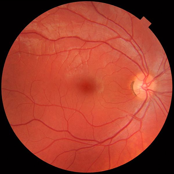
English: Fundus photograph of the right eye, showing a fundus with no sign of disease or pathology. It is seen from front so that left in each image is to the person's right. The gaze is into the camera, so the macula is in the center of the image, and the optic disk is located towards the nose (right in image). The optic disk has some pigmentation at the perimeter of the lateral side, which is considered non-pathological.
Veins are darker and slightly wider than corresponding arteries. Major nerve pathways are seen as white striped patterns radiating from the optic disk. In addition, there are also lighter areas close to larger vessels seen mainly at upper left in the image (person's upper right), which is regarded as a normal finding in younger people. Photo is taken at Gävle Hospital in Sweden in 2012 on a healthy 25-year old male volunteer. |
| Tarih | |
| Kaynak | Yükleyenin kendi çalışması |
| Yazar |
When using this image in external works, it may be cited as:
or
|
| Diğer sürümler |
 |
Lisanslama
Ben, bu işin telif sahibi, burada işi aşağıdaki lisans altında yayımlıyorum:
| Bu dosya Creative Commons Evrensel Kamu Malı İthafı altındadır. | |
| Bu çalışmayı oluşturan kişi bu senet ile eser hakkında tüm dünya çapında telif hakkı yasaları kapsamında, yasalar tarafından izin verilen ölçülerde ve diğer benzer tüm haklarından feragat etmiş ve kamu malı olarak nitelendirmiştir. Siz bu çalışmayı ve eseri hiç bir izin almadan ticari amaçlar da dahil olmak üzere kopyalayabilir, değiştirebilir ve serbestçe dağıtabilirsiniz.
|
Dosya geçmişi
Dosyanın herhangi bir zamandaki hâli için ilgili tarih/saat kısmına tıklayın.
| Tarih/Saat | Küçük resim | Boyutlar | Kullanıcı | Yorum | |
|---|---|---|---|---|---|
| güncel | 12.49, 21 Mart 2012 | | 1.411 × 1.411 (248 KB) | wikimediacommons>Mikael Häggström |
Dosya kullanımı
Aşağıdaki sayfa bu dosyayı kullanmaktadır: